Photoacoustic and Ultrasound Imaging Laboratory

光聲暨超音波影像實驗室

Research

Research Projects:


  • Ultrafast Doppler


  • Ultrasound-mediated drug delivery

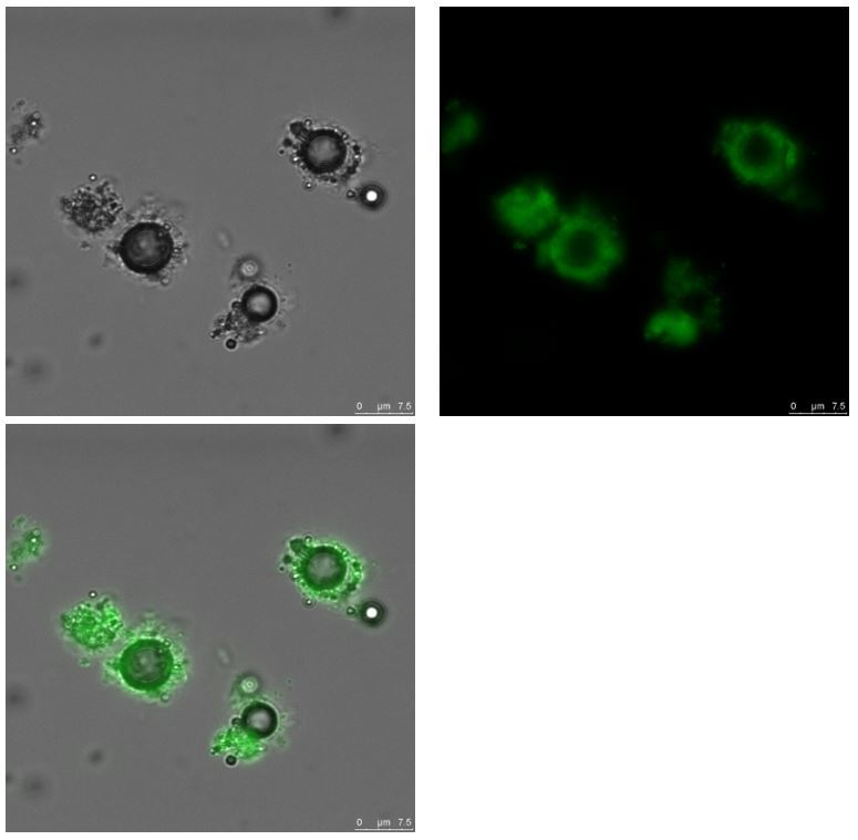
    Boron Neutron Capture Therapy (BNCT) is a new tumor targeting therapy for treating locally invasive malignant tumors such as recurrent head and neck cancer, and primary brain tumor. BNCT is based on thermal neutron irradiation interacting with enriched 10B in the tumor region. The thermal neutron capture reaction (10B(n,α)7Li), gives rise to two high linear energy transfer (LET) particles that release all their energy in a path of the order of a cell diameter. If tumor uptake higher concentration of boron containing drug compared to normal cells, the tumor cells are selectively/targeting damaged with a substantial sparing of the healthy surrounding tissue, and with lower side effects to the patient. The major challenge in the development of boron delivery agents has been the requirement for selective tumor targeting. In the preliminary study, the silicon nanoparticles were synthesized with Ouzo effect and the fluoresceinamine (FA) molecules were embedded within particles. The average size and zeta potential are 121.7 1.1 nm and +49.2 4.7 mV, respectively. The FA was controlled released with high intensity focused ultrasound (HIFU) irradiation of 75 watts and 3 mins in the in-vitro test. This ultrasound-mediated drug delivery system will be further applied to in-vivo experiments, and tumor will be even treated with boron neutron capture therapy.

  • Microscopic image of FA-SiNp-MBs                            Ultrasound-mediated drug release


  • Shear Wave Elastography

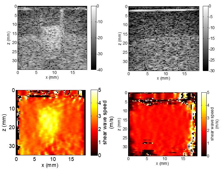
    • Ultrasound Shear Wave Imaging

      heterogeneous                               homogeneous


    • Shear wave tracking

      heterogeneous

      homogeneous 

    • Optical Coherence Elastography

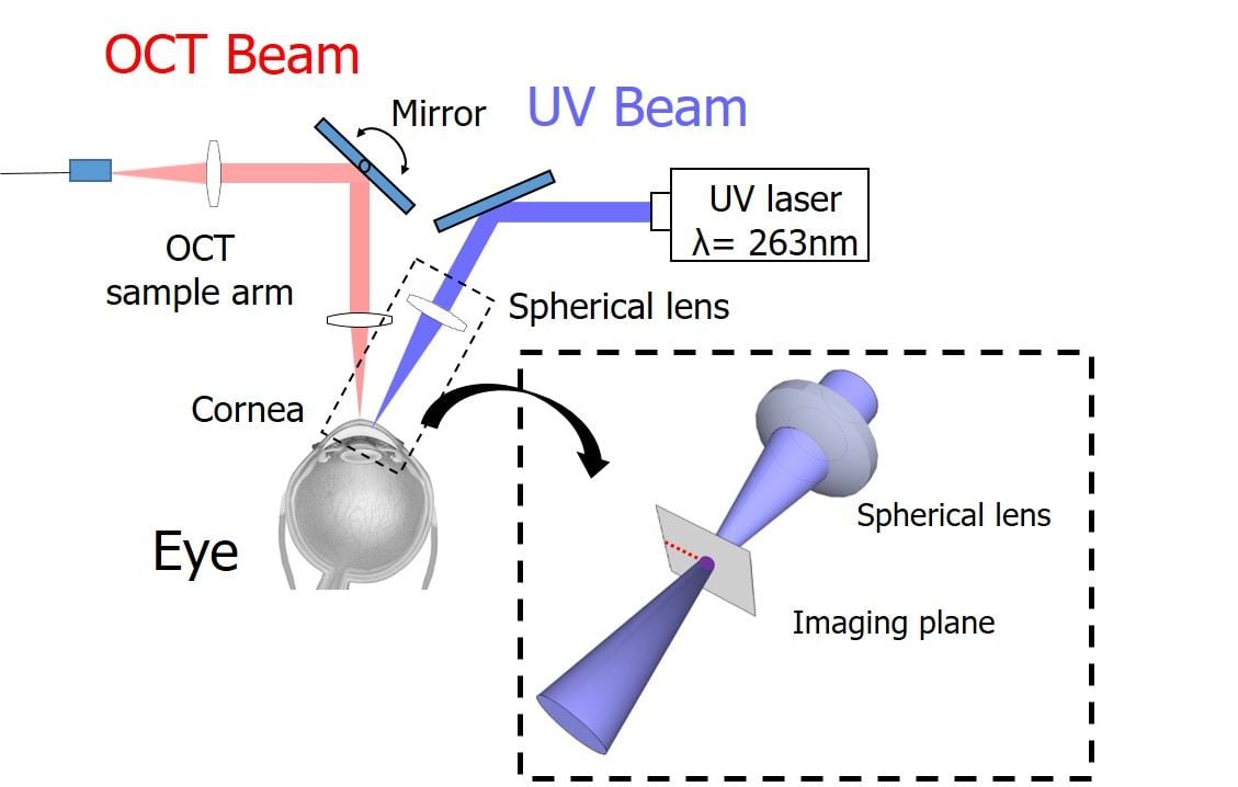
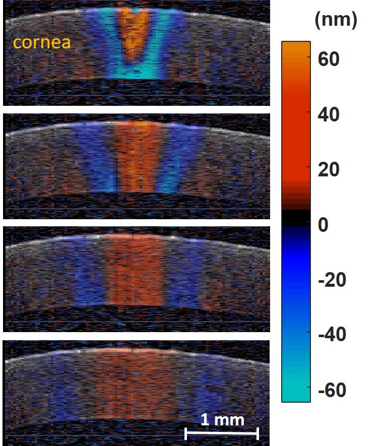
      Corneal biomechanics is critical for early diagnosis, optimal management of corneal diseases (e.g keratoconus) and for predicting the risks of surgical intervention of healthy corneas, such as post-LASIK ectasia. In this study, elasticity measurements of ex-vivo porcine corneal tissue are demonstrated with non-contact optical coherence elastography (OCE) by integrating ultraviolet (UV) laser pulse excitation and high speed phase-sensitive optical coherence tomography (PhS-OCT). The results respond linearly to intraocular pressure (IOP) (from 4-28 mmHg) based on the measurement of and group velocity (R2= 0.93) of propagating waves in the cornea launched by a single UV laser pulse. In addition, the 2-D elastic map of corneal tissue are obtained. This clinically-translatable OCE system can potentially generate personalized biomechanical models to help early diagnosis, and continued monitoring of keratoconus, pre-refractive screening for risks of ectasia, and treatment monitoring.

  • Computer-aided Diagnosis

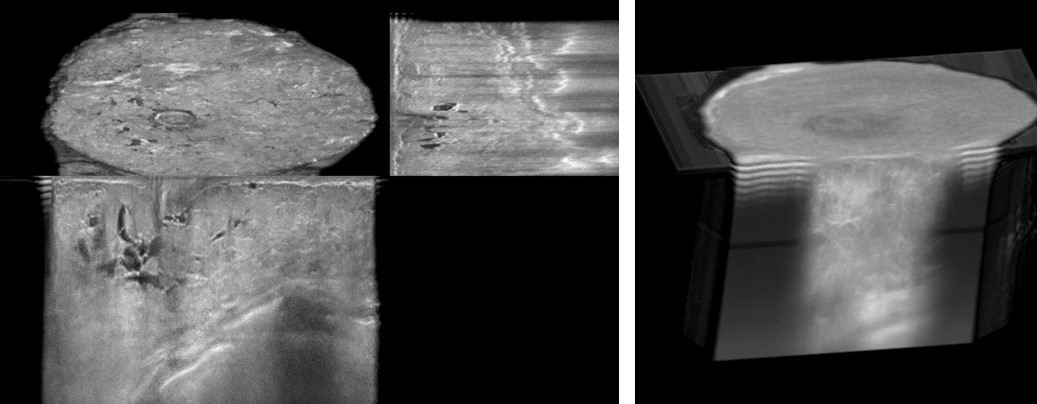
    Breast cancer is the most common cancer in women, and the prevalence rate is increasing in recent years. Many studies have shown that higher breast density is highly correlated to higher risk for breast cancer. Mammography is the main screening imaging tool to access breast density and then detect early breast cancer. However, the scanning procedure makes patients uncomfortable due to breast compression, and patients are exposed to ionizing radiation. In addition, tissue overlap is the major limitation for diagnosis with 2-D mammography. Automated breast volume scanner (ABVS) is a new imaging system with automated ultrasound breast scan and has potential to have 3-D imaging data for breast density analysis. In this study, we proposed to calculate breast density with volume data from ABVS. To approach this goal, we suggest a semi-automatic image process by using fuzzy C-means classifier to extract the fibro-glandular tissues from the acquired images. From this preliminary result, we found that breast density analysis with the ABVS 3-D images can provide a potential computer-aided diagnosis for breast screening and will be demonstrated the efficacy with more clinical cases.


Skip to toolbar